Unraveling Altered Parenchymal Echotexture: Unlocking Medical Mysteries
Altered parenchymal echotexture refers to changes in the normal appearance of an organ or tissue on an ultrasound examination, often indicating underlying disease or abnormality. The echotexture, or pattern of echoes, produced by ultrasound waves bouncing off the tissue can provide valuable information about its structure and composition. When the echotexture is altered, it may suggest the presence of inflammation, fibrosis, fatty infiltration, or other pathological processes.
Altered parenchymal echotexture plays a crucial role in medical diagnostics, as it can help identify a wide range of conditions. For example, in the liver, altered echotexture can indicate cirrhosis, hepatitis, or fatty liver disease. In the kidneys, it can suggest chronic kidney disease or pyelonephritis. In the thyroid gland, it can help diagnose thyroid nodules or autoimmune thyroiditis. By detecting and interpreting altered parenchymal echotexture, physicians can make informed decisions about further diagnostic tests, treatments, and patient management.
The evaluation of altered parenchymal echotexture is an essential component of ultrasound examinations, and it requires specialized training and experience. Radiologists and sonographers use their knowledge of normal and abnormal echotexture patterns to identify and characterize tissue abnormalities, contributing to accurate diagnoses and effective patient care.
What is Altered Parenchymal Echotexture?
Altered parenchymal echotexture refers to changes in the normal appearance of an organ or tissue on an ultrasound examination. The term "echotexture" describes the pattern of echoes produced by ultrasound waves bouncing off the tissue, and alterations in this pattern can indicate underlying disease or abnormality.
- Definition: Changes in the normal appearance of an organ or tissue on ultrasound.
- Causes: Inflammation, fibrosis, fatty infiltration, and other pathological processes.
- Importance: Can help identify a wide range of medical conditions.
- Diagnostic value: Used to evaluate the liver, kidneys, thyroid gland, and other organs.
- Interpretation: Requires specialized training and experience.
- Examples: Cirrhosis, hepatitis, fatty liver disease, chronic kidney disease, pyelonephritis, thyroid nodules.
- Clinical implications: Can guide further diagnostic tests, treatments, and patient management.
- Future directions: Ongoing research to improve the accuracy and reliability of echotexture evaluation.
In summary, altered parenchymal echotexture is a valuable tool for medical diagnostics, providing insights into the health of various organs and tissues. By detecting and interpreting these changes, physicians can make informed decisions about patient care and improve overall health outcomes.
Definition
Altered parenchymal echotexture is defined as changes in the normal appearance of an organ or tissue on ultrasound. This definition highlights the fundamental characteristic of altered parenchymal echotexture, which is a deviation from the expected echotexture pattern of a healthy organ or tissue. The echotexture, or pattern of echoes, produced by ultrasound waves bouncing off the tissue provides valuable information about its structure and composition. When the echotexture is altered, it may suggest the presence of underlying disease or abnormality.
The definition of altered parenchymal echotexture is crucial for understanding its significance in medical diagnostics. By identifying changes in the normal echotexture pattern, physicians can gain insights into the health of various organs and tissues. For example, in the liver, altered echotexture can indicate cirrhosis, hepatitis, or fatty liver disease. In the kidneys, it can suggest chronic kidney disease or pyelonephritis. In the thyroid gland, it can help diagnose thyroid nodules or autoimmune thyroiditis.
Understanding the definition of altered parenchymal echotexture is essential for interpreting ultrasound examinations and making informed decisions about patient care. Radiologists and sonographers rely on their knowledge of normal and abnormal echotexture patterns to identify and characterize tissue abnormalities, contributing to accurate diagnoses and effective patient management.
Causes
Altered parenchymal echotexture is often caused by underlying pathological processes that affect the structure and composition of an organ or tissue. These pathological processes can include inflammation, fibrosis, fatty infiltration, and a range of other abnormalities.
- Inflammation: Inflammation is a natural response to injury or infection, characterized by the infiltration of immune cells and fluid into the affected tissue. This influx of cells and fluid can alter the echotexture of the tissue, making it appear more heterogeneous and less distinct.
- Fibrosis: Fibrosis is the excessive formation of fibrous connective tissue in response to chronic injury or inflammation. This can lead to the replacement of normal tissue with scar tissue, which has a different echotexture than healthy tissue. Fibrosis can make the echotexture appear more coarse and irregular.
- Fatty infiltration: Fatty infiltration occurs when fat cells accumulate in an organ or tissue that normally does not contain a significant amount of fat. This can alter the echotexture of the tissue, making it appear more hyperechoic (brighter) and less homogeneous.
- Other pathological processes: In addition to inflammation, fibrosis, and fatty infiltration, a variety of other pathological processes can also cause altered parenchymal echotexture. These include tumors, cysts, abscesses, and vascular malformations. Each of these conditions can produce characteristic changes in the echotexture of the affected tissue.
Understanding the causes of altered parenchymal echotexture is crucial for interpreting ultrasound examinations and making informed decisions about patient care. By identifying the underlying pathological process, physicians can determine the appropriate course of action, whether it involves further diagnostic tests, treatment, or monitoring.
Importance
Altered parenchymal echotexture is of significant importance in medical diagnostics, as it can help identify a wide range of medical conditions affecting various organs and tissues. By detecting and interpreting changes in the normal echotexture pattern, physicians can gain valuable insights into the health of these organs and tissues, leading to more accurate diagnoses and appropriate patient management.
- Early disease detection: Altered parenchymal echotexture can sometimes indicate the presence of disease even before symptoms appear. For example, in the liver, altered echotexture can be an early sign of fatty liver disease, which, if left untreated, can lead to liver damage and scarring.
- Differential diagnosis: Altered parenchymal echotexture can help differentiate between different medical conditions with similar symptoms. For instance, in the kidneys, altered echotexture can help distinguish between acute pyelonephritis and chronic kidney disease, which have similar symptoms but require different treatments.
- Monitoring disease progression: Serial ultrasound examinations can be used to monitor the progression of certain medical conditions by assessing changes in parenchymal echotexture. For example, in patients with chronic liver disease, monitoring changes in liver echotexture can help assess the response to treatment and disease progression.
- Guiding biopsies and other procedures: Altered parenchymal echotexture can guide biopsies and other interventional procedures by providing a roadmap of the affected tissue. This can increase the accuracy and safety of these procedures, especially when dealing with small or deep-seated lesions.
In summary, the importance of altered parenchymal echotexture lies in its ability to help identify a wide range of medical conditions, facilitate differential diagnosis, monitor disease progression, and guide interventional procedures. By leveraging this information, physicians can make more informed decisions about patient care, leading to improved health outcomes.
Diagnostic value
Altered parenchymal echotexture has significant diagnostic value, as it can be used to evaluate a wide range of organs and tissues throughout the body. By assessing changes in the normal echotexture pattern, physicians can gain valuable insights into the health of these organs and tissues, leading to more accurate diagnoses and appropriate patient management.
- Liver: Altered parenchymal echotexture can help identify a variety of liver conditions, including cirrhosis, hepatitis, and fatty liver disease. For example, in cirrhosis, the liver echotexture may appear coarse and irregular due to the replacement of normal liver tissue with scar tissue.
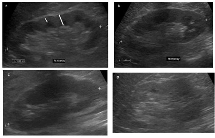
- Kidneys: In the kidneys, altered parenchymal echotexture can indicate chronic kidney disease, pyelonephritis, and other renal abnormalities. For instance, in chronic kidney disease, the kidney echotexture may appearbrighter and less distinct due to the loss of
- Thyroid gland: Altered parenchymal echotexture can help diagnose thyroid nodules, autoimmune thyroiditis, and other thyroid conditions. For example, in thyroid nodules, the echotexture may appear heterogeneous and contain areas of increased or decreased echogenicity.
- Other organs: Altered parenchymal echotexture can also be used to evaluate the spleen, pancreas, lymph nodes, and other organs. Each organ has its own characteristic echotexture pattern, and deviations from this pattern can indicate underlying disease or abnormality.
Overall, the diagnostic value of altered parenchymal echotexture lies in its ability to provide detailed information about the structure and composition of various organs and tissues. By detecting and interpreting changes in echotexture, physicians can make informed decisions about further diagnostic tests, treatments, and patient management, ultimately leading to improved health outcomes.
Interpretation
The interpretation of altered parenchymal echotexture requires specialized training and experience, as it involves the recognition and characterization of subtle changes in the echotexture pattern. This expertise is crucial for accurate diagnosis and patient management.
Understanding the connection between altered parenchymal echotexture and the need for specialized training is essential for several reasons:
- Complexity of echotexture patterns: Echotexture patterns can vary widely depending on the organ or tissue being examined, the underlying pathological process, and the ultrasound equipment used. Specialized training helps sonographers and radiologists develop the skills necessary to recognize and differentiate between normal and abnormal echotexture patterns.
- Subtle changes in echogenicity: Altered parenchymal echotexture is often characterized by subtle changes in echogenicity, which can be difficult to detect and interpret without proper training. Specialized training enhances the ability to identify these subtle changes and determine their significance.
- Correlation with clinical findings: The interpretation of altered parenchymal echotexture should be done in conjunction with the patient's clinical history and other diagnostic findings. Specialized training helps healthcare professionals integrate all available information to make accurate diagnoses and determine appropriate patient management.
In summary, the interpretation of altered parenchymal echotexture is a complex task that requires specialized training and experience. This expertise is essential for accurate diagnosis and patient management, as it enables healthcare professionals to recognize and characterize subtle changes in echotexture patterns and correlate them with clinical findings.
Examples
The examples providedcirrhosis, hepatitis, fatty liver disease, chronic kidney disease, pyelonephritis, and thyroid nodulesare medical conditions that can manifest as altered parenchymal echotexture on ultrasound examinations. Understanding this connection is crucial for accurate diagnosis and patient management.
Altered parenchymal echotexture, as discussed earlier, refers to changes in the normal appearance of an organ or tissue on ultrasound. These changes are often caused by underlying pathological processes, such as inflammation, fibrosis, fatty infiltration, and others. The examples provided represent various conditions associated with specific pathological processes that lead to altered echotexture patterns.
For instance, in cirrhosis, a chronic liver disease, the liver's normal echotexture is altered due to the replacement of healthy liver tissue with scar tissue. This scarring process, known as fibrosis, results in a coarse and irregular echotexture. Similarly, in chronic kidney disease, the kidneys' echotexture may appear brighter and less distinct due to the loss of normal kidney tissue and its replacement with fibrotic tissue.
By recognizing the connection between altered parenchymal echotexture and specific medical conditions, healthcare professionals can effectively interpret ultrasound findings and make informed decisions about further diagnostic tests, treatments, and patient management. This understanding forms the cornerstone of accurate and timely medical care.
Clinical implications
Altered parenchymal echotexture has significant clinical implications, as it can guide further diagnostic tests, treatments, and patient management. Understanding this connection is crucial for healthcare professionals to make informed decisions and provide optimal care to patients.
- Diagnostic implications: Altered parenchymal echotexture can indicate the presence of underlying medical conditions, prompting further diagnostic tests to confirm the diagnosis. For example, in the liver, altered echotexture may suggest cirrhosis or fatty liver disease, leading to additional tests such as liver function tests or a liver biopsy.
- Treatment implications: The identification of altered parenchymal echotexture can influence treatment decisions. For instance, in the kidneys, altered echotexture may indicate chronic kidney disease, which may require specific medications or lifestyle modifications to manage the condition.
- Patient management implications: Monitoring changes in parenchymal echotexture over time can provide valuable information about the progression of a disease or the response to treatment. This information can guide patient management decisions, such as adjusting medications or scheduling follow-up examinations.
- Prognostic implications: In some cases, altered parenchymal echotexture can provide prognostic information. For example, in thyroid nodules, the echotexture can help assess the likelihood of malignancy, influencing decisions about further evaluation or treatment.
By understanding the clinical implications of altered parenchymal echotexture, healthcare professionals can effectively interpret ultrasound findings, make informed decisions about patient care, and improve overall patient outcomes.
Future directions
The exploration of "Future directions: Ongoing research to improve the accuracy and reliability of echotexture evaluation" is closely connected to the understanding of "what is altered parenchymal echotexture." This connection stems from the fact that accurate and reliable echotexture evaluation is crucial for effective disease diagnosis and patient management.
- Advanced imaging techniques: Research is ongoing to develop and refine advanced imaging techniques, such as elastography and shear wave elastography, which can provide additional information about tissue stiffness and elasticity. This can enhance the accuracy of echotexture evaluation, particularly in differentiating between benign and malignant lesions.
- Artificial intelligence (AI): AI algorithms are being developed to assist in the interpretation of echotexture patterns. These algorithms can analyze large datasets of ultrasound images and identify subtle changes that may be difficult for the human eye to detect. This can improve the reliability and consistency of echotexture evaluation.
- Standardization of echotexture assessment: Researchers are working to establish standardized protocols for echotexture assessment, ensuring that it is performed consistently across different ultrasound machines and operators. This standardization can improve the comparability of echotexture findings and facilitate the development of more accurate and reliable diagnostic criteria.
- Integration with other imaging modalities: Research is also exploring the integration of echotexture evaluation with other imaging modalities, such as computed tomography (CT) and magnetic resonance imaging (MRI). This can provide a more comprehensive view of the tissue being examined and improve the accuracy of diagnosis.
The ongoing research in these areas aims to improve the accuracy and reliability of echotexture evaluation, which will ultimately lead to better patient care and outcomes. By refining imaging techniques, utilizing AI, standardizing assessment protocols, and integrating with other imaging modalities, the field of echotexture evaluation is continuously advancing, enhancing its role in medical diagnostics and patient management.
Frequently Asked Questions About Altered Parenchymal Echotexture
This section presents answers to frequently asked questions about altered parenchymal echotexture, providing a deeper understanding of its significance and implications.
Question 1: What exactly is altered parenchymal echotexture?Altered parenchymal echotexture refers to changes in the normal appearance of an organ or tissue on an ultrasound examination. These changes are often caused by underlying disease or abnormality, and their detection can aid in diagnosis and patient management.
Question 2: How important is altered parenchymal echotexture in medical diagnostics?Altered parenchymal echotexture plays a crucial role in medical diagnostics, as it can help identify a wide range of conditions affecting various organs and tissues. By detecting and interpreting these changes, physicians gain valuable insights into the health of these organs and tissues, leading to more accurate diagnoses.
Question 3: Which organs can be evaluated using altered parenchymal echotexture?Altered parenchymal echotexture has diagnostic value in evaluating a variety of organs, including the liver, kidneys, thyroid gland, spleen, pancreas, and lymph nodes. Each organ has its own characteristic echotexture pattern, and deviations from this pattern may indicate underlying disease or abnormality.
Question 4: How do healthcare professionals interpret altered parenchymal echotexture?The interpretation of altered parenchymal echotexture requires specialized training and experience. Healthcare professionals, such as sonographers and radiologists, are trained to recognize and characterize subtle changes in echotexture patterns, which can provide valuable information about the underlying tissue structure and composition.
Question 5: What are the clinical implications of altered parenchymal echotexture?Altered parenchymal echotexture has significant clinical implications, as it can guide further diagnostic tests, treatments, and patient management. By understanding the connection between altered echotexture and specific medical conditions, healthcare professionals can make informed decisions about the best course of action for each patient.
Question 6: Is there ongoing research to improve the accuracy and reliability of echotexture evaluation?Yes, ongoing research aims to improve the accuracy and reliability of echotexture evaluation. Researchers are exploring advanced imaging techniques, artificial intelligence (AI) algorithms, standardized assessment protocols, and integration with other imaging modalities to enhance the effectiveness of echotexture evaluation in medical diagnostics and patient care.
In summary, altered parenchymal echotexture is a valuable tool in medical diagnostics, providing insights into the health of various organs and tissues. Its interpretation requires specialized training and experience, and it has significant clinical implications. Ongoing research is focused on improving the accuracy and reliability of echotexture evaluation, further enhancing its role in patient care.
Transition to the next article section...
Tips for Understanding Altered Parenchymal Echotexture
1
2
3
4
5
6
Conclusion
Altered parenchymal echotexture is a valuable tool in medical diagnostics, providing insights into the health of various organs and tissues. Its interpretation requires specialized training and experience, and it has significant clinical implications. Ongoing research aims to improve the accuracy and reliability of echotexture evaluation, further enhancing its role in patient care.
By understanding the significance of altered parenchymal echotexture, healthcare professionals can effectively utilize ultrasound examinations to detect and characterize tissue abnormalities, leading to more precise diagnoses and appropriate patient management. This ultimately contributes to improved patient outcomes and a better understanding of various medical conditions.
Unveiling The Guardians Behind Figure Skating Prodigy: Alexandra Trusova's Parents
Uncover The Extraordinary Journey Of Paula Jai Parker: A Biography Unveiled
Caprice Herjavec: Uncovering The Journey Of Robert Herjavec's Accomplished Daughter
High resolution ultrasound of liver (A) shows diffusely altered
Primary Highgrade Peripheral Tcell Lymphoma of the Testis Clinically